This higher tier covers Higher Brain Scanning and Vision Defects within Nervous System for GCSE Biology. Topic 2: Nervous System It is section 15 of 18 in this topic. This section is most useful once the core foundation idea is secure, because it adds the detail that pushes answers higher.
Higher Brain Scanning and Vision Defects
Brain scanning techniques
Scientists use several scanning methods to study the brain:
- CT scans — use X-rays to build up a detailed image of brain structure. Useful for detecting tumours or bleeding.
- PET scans — detect radioactive tracers to show which brain areas are most active. Useful for mapping brain function.
- MRI scans — use strong magnetic fields to produce detailed images of soft tissue. No radiation involved.
These techniques have improved our understanding, but there are limitations: we still do not fully understand how complex behaviours, emotions, and memory work. Treating brain damage remains very difficult because neurones rarely regenerate.
Vision defects
| Defect | Cause | Effect | Correction |
|---|---|---|---|
| Myopia (short-sightedness) | Eyeball too long or lens too curved | Image focuses in front of retina — distant objects blurry | Concave (diverging) lens |
| Hyperopia (long-sightedness) | Eyeball too short or lens too flat | Image focuses behind retina — close objects blurry | Convex (converging) lens |
Both can also be treated with laser eye surgery (reshapes the cornea) or replacement lenses.
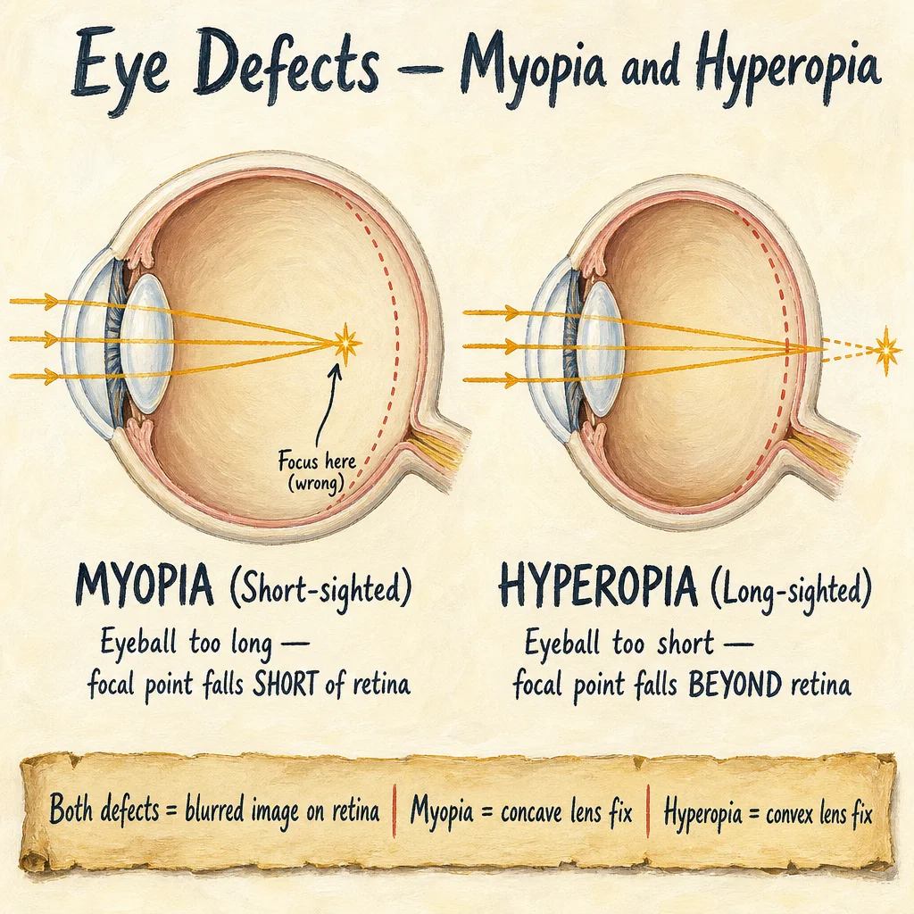
Figure 3: How myopia and hyperopia cause incorrect focusing of light on the retina

Figure 4: Corrective lenses redirect light to focus exactly on the retina
Practice questions for Nervous System
What are the two organs that make up the central nervous system (CNS)?
Explain how a signal is transmitted across a synapse from one neurone to the next.