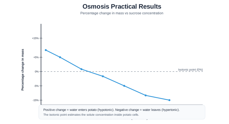
Typical results showing relationship between sucrose concentration and percentage change in mass
Interpreting Results
- Positive percentage change: Water moved into potato — the external solution was more dilute (higher water concentration) than the potato cells
- Negative percentage change: Water moved out of potato — the external solution was more concentrated (lower water concentration) than the potato cells
- Zero percentage change: The solution and potato cells had equal concentrations — no net movement of water
- Finding the equal point: Read from the graph where the line crosses the x-axis — this tells you the concentration of solutes inside the potato cells
Quick Check: A student uses iodine solution to stain an onion cell slide. What colour would you expect the cell wall region to appear, and why does staining help?
The cell wall (and any starch present) would appear dark blue-black after iodine staining. Staining is necessary because cell structures are mostly colourless and transparent without dye — they would be almost invisible under the microscope. The iodine binds to the cell wall components, adding contrast so the structure can be clearly observed and drawn.
This diagram covers Osmosis Results and Analysis within Cell Biology Practical Investigations for GCSE Biology. Comprehensive practical skills, experimental design, data analysis, microscopy techniques, and scientific methodology in cell biology It is section 9 of 18 in this topic. Focus on the labels, the relationships between parts, and the explanation that turns the diagram into an exam-ready answer.
Practice questions for Cell Biology Practical Investigations
When using a light microscope to observe cells, which objective lens should be used first?
Describe how to focus a light microscope on a specimen.