The City Inside Every Cell
🔬 The City Inside Every Cell
Think of a cell as a tiny, self-sufficient city. It has a control centre giving instructions (the nucleus), power stations generating energy (mitochondria), factories building essential products (ribosomes), and a postal sorting office (the Golgi apparatus). Every organelle has a job, and when they all work together, the cell stays alive.
Understanding organelles is one of the most important foundations of GCSE Biology. Questions about organelle structure and function appear in virtually every unit — from respiration and photosynthesis to genetics and infection. Get these right and you have a head start across the whole course.
A typical plant cell. Tap any label to explore its function.
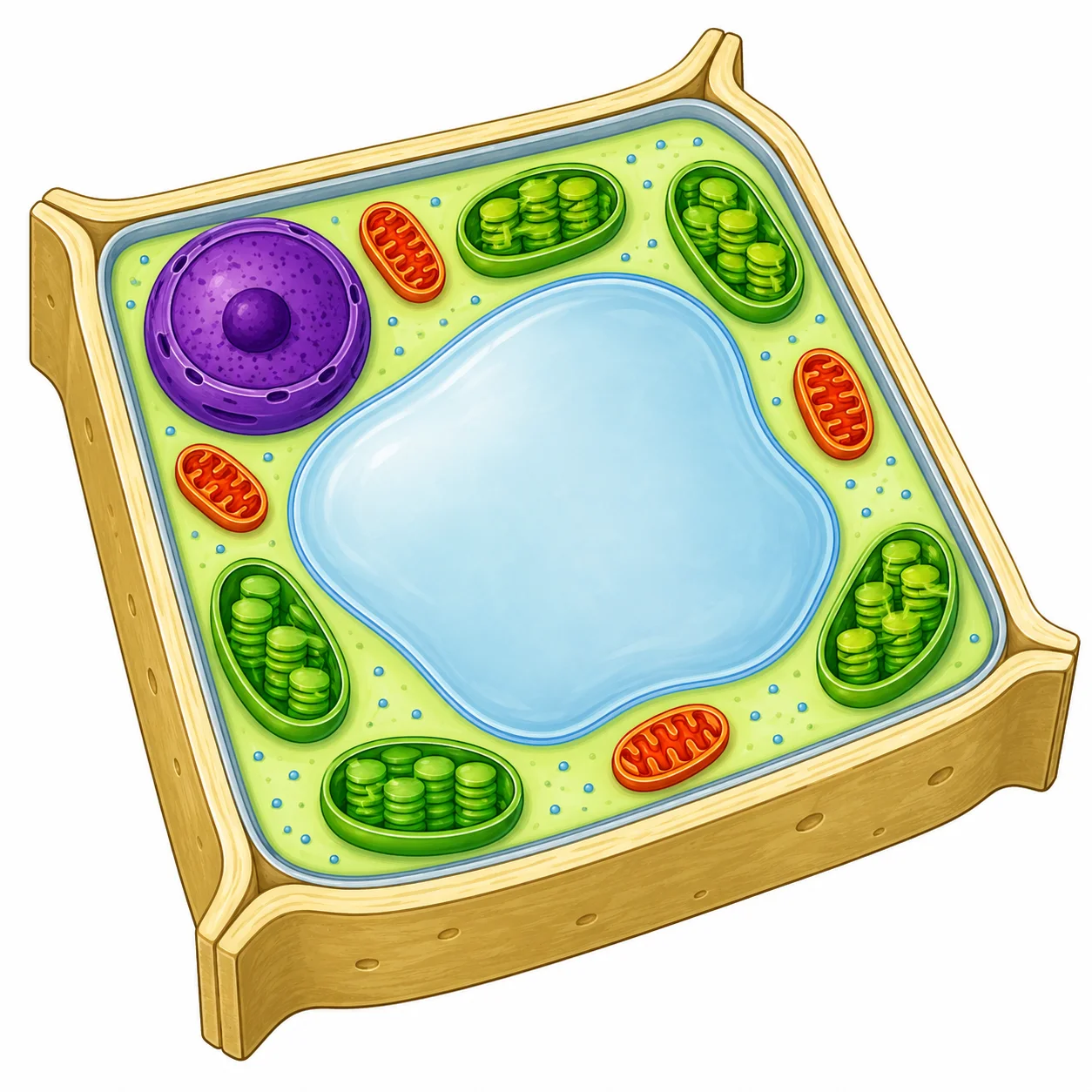
- 1Cell wall — Rigid outer layer made of cellulose. Provides structural support.
- 2Nucleus — Contains DNA. Controls all cell activities — the cell's control centre.
- 3Chloroplast — Site of photosynthesis. Contains chlorophyll which absorbs light.
- 4Permanent vacuole — Large fluid-filled sac that maintains turgor pressure in the cell.
- 5Mitochondrion — Site of aerobic respiration — releases energy. Present in plant cells too.
- 6Cell membrane — Controls what enters and leaves the cell. Sits just inside the cell wall.
Figure 2: A typical plant cell. Tap any label to explore its function.
Biology glossary
- What is the formula for calculating magnification?
- Magnification = image size ÷ real size (both measurements must be converted to the same units before dividing). For example, if an image measures 6000 μm across and the real object measures 2 μm across, the magnification is 6000 ÷ 2 = ×3000.
- What is the difference between magnification and resolution?
- Magnification: how many times larger the image is than the real object. Resolution: the ability to distinguish two nearby points as separate. A high-resolution image shows fine detail; high magnification of a low-resolution image just produces a blurry enlargement.
Everything on this page — the cristae inside mitochondria, the grana inside chloroplasts, even the existence of ribosomes — was discovered using microscopes. To use microscope images properly, you need to understand resolution, magnification, units, and when to estimate rather than count exactly.
Earn the mark scheme marks
💡 Memory Aid: The Cell as a Factory
Imagine the cell is a factory. Each organelle has a job — match each one to its factory role:
| Organelle | Factory Role | What It Actually Does |
|---|---|---|
| Nucleus | 🏢 Boss's office | Contains DNA — controls all cell activities |
| Cell membrane | 🚧 Security gate | Controls what enters and leaves the cell |
| Cytoplasm | 🏭 Factory floor | Where most chemical reactions take place |
| Mitochondria | ⚡ Power station | Site of aerobic respiration — transfers energy |
| Ribosomes | 🔧 Assembly line | Where proteins are made (protein synthesis) |
Plant-Only Extras: "WCV" — Wall, Chloroplast, Vacuole
- Cell wall = outer fence (rigid cellulose — supports and protects)
- Chloroplast = solar panels (absorbs light for photosynthesis)
- Permanent vacuole = storage warehouse (filled with cell sap — keeps plant rigid)
Quick Trick for Exams
If asked "Which organelle...?" — mitochondria is almost always the answer when the question mentions energy, active cells, or respiration. Ribosomes is the answer when the question mentions protein or growth.
Now try it yourself
Quiz · Question 1 of 12
Scientists studying mitochondria use electron microscopes rather than light microscopes. Which statement correctly explains why electron microscopes are more useful for studying cell ultrastructure?
Tap an answer to check it