The World Invisible to the Naked Eye
🔬 The World Invisible to the Naked Eye
For thousands of years, humans could only see what their eyes allowed them to see. Then in the late 1500s, Dutch lens makers began creating the first microscopes, opening up an entirely new world of discovery. When Robert Hooke peered through his microscope at a slice of cork in 1665, he saw tiny box-like structures that he called "cells" - launching the field of cell biology.
Today, microscopes are essential tools in biology, allowing us to explore the intricate structures that make up all living things. From the delicate organelles inside a single cell to the complex tissues of multicellular organisms, microscopy reveals the hidden architecture of life.
Light microscope — click each dot to explore the parts and how they work
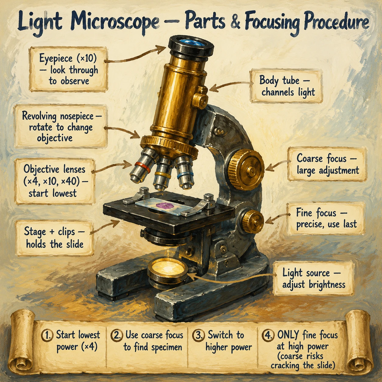
- 1Eyepiece lens (×10) —
- 2Revolving nosepiece —
- 3Objective lenses —
- 4Stage with clips —
- 5Coarse focus knob —
- 6Fine focus knob —
- 7Light source —
Light microscope — click each dot to explore the parts and how they work
Biology glossary
- What is resolution?
- Resolution is the ability to distinguish between two separate points that are close together. It determines how clear and detailed an image is.
- What is magnification?
- Magnification is how many times larger an image appears compared to the actual object. It tells us how much bigger something looks through a microscope.
Earn the mark scheme marks
Memory Aids: Microscopy
The MIA Triangle for Magnification Calculations
Remember the three quantities with the word MIA:
- M = Magnification
- I = Image size (what you measure on the image)
- A = Actual size (real size of the object)
Draw a triangle with I on top, and A x M on the bottom. Cover the letter you want to find:
- Cover M: Magnification = I / A
- Cover I: Image size = A x M
- Cover A: Actual size = I / M
Light vs Electron: "LED vs BEAM"
To remember what each type uses:
- Light microscope uses Light (LED bulb) — Living specimens possible
- Electron microscope uses Electron beam — Everything must be dead
TEM vs SEM: "T is for Through, S is for Surface"
- TEM — electrons pass Through the specimen — gives 2D internal images
- SEM — electrons Scan the surface — gives 3D external images
Unit Conversions: "Move the decimal 3 places"
Each step in the unit ladder is x1000:
- mm to μm: multiply by 1000 (move decimal 3 places right)
- μm to nm: multiply by 1000 (move decimal 3 places right)
- nm to μm: divide by 1000 (move decimal 3 places left)
- μm to mm: divide by 1000 (move decimal 3 places left)
Quick Check: Under a light microscope, a cell image measures 15 mm. The microscope is set to x300 total magnification. Calculate the actual size of the cell. Give your answer in micrometres (μm).
Actual size = Image size / Magnification = 15 mm / 300 = 0.05 mm. Convert to μm: 0.05 mm x 1000 = 50 μm. The actual size of the cell is 50 μm.
Quick Check: A student wants to study the internal structure of a ribosome, which has a diameter of approximately 20 nm. Explain why a light microscope cannot be used for this purpose, and state which type of microscope would be needed.
A light microscope has a maximum resolution of approximately 200 nm — it cannot distinguish two points closer than 200 nm. A ribosome is only 20 nm in diameter, which is 10 times smaller than this resolution limit. Even at maximum magnification, the ribosome would be invisible or blurred because the wavelength of light is too large to resolve it. A Transmission Electron Microscope (TEM) would be needed, as it has a resolution of 0.05 nm — far smaller than a ribosome — and can reveal internal cellular structures in detail.
Quick Check: A scientist is choosing between a light microscope and an electron microscope to study white blood cells engulfing bacteria in real-time. Which should they choose, and why? What limitation does the alternative microscope have for this purpose?
The scientist should choose a light microscope. Light microscopes can view living specimens in real-time, allowing the process of phagocytosis (white blood cells engulfing bacteria) to be observed as it happens. An electron microscope cannot be used for this purpose because the preparation process kills the specimen — specimens must be completely dehydrated and placed in a vacuum. An electron microscope can only show a snapshot of a dead, fixed cell, not a dynamic live process.
Now try it yourself
Quiz · Question 1 of 25
What is magnification?
Tap an answer to check it
This topic in real past papers
Every real exam question we've found on microscopy, with a full worked answer.