Figure 8 is a CT image of a head with a brain tumour. PET scans are another way to find tumours, and monoclonal antibodies can be used to locate blood clots.
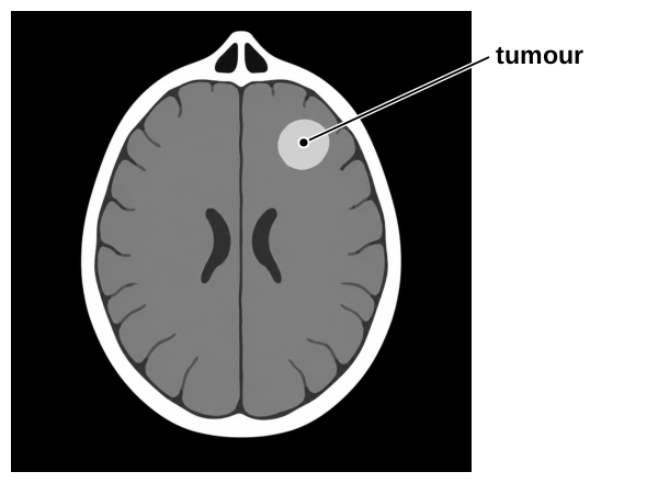
Why does a CT scan show up a tumour?
Our own questions on this topic, marked straight away, so you see what to revise. Free now.
Show model answer for 07.1Hide model answer for 07.1
A CT scan sends X-rays through the head. The tumour absorbs the X-rays.
This happens because a tumour is a dense mass of cells, so it looks different from the tissue around it.
How the marks are earned
- Three linked ideas, of which two earn the marks: X-rays are used; the tumour absorbs them; a tumour is a mass of cells, or is dense (either earns it).
- Saying that tumours absorb the radiation is credited.
- The statement tumours absorb X-rays, alone, earns both marks.
Why it scores
These are the first two points. The single idea of a tumour absorbing X-rays earns both marks.
This is the reason, and it explains why the tumour can be seen on the image.
Revise Cancer and Cell Division ControlWhich region of the brain deals with sight? Name it.
See how you would score on Nervous SystemShow model answer for 07.2Hide model answer for 07.2
The cerebral cortex, which is part of the cerebrum.
How the marks are earned
- Cerebrum, cerebral hemispheres or cerebral cortex.
- Also credited as printed: hippocampus; occipital lobe; cerebellum; frontal lobe.
A PET scan starts with an injection of radioactive glucose. Why is it injected?
See how you would score on Cancer and Cell Division ControlShow model answer for 07.3Hide model answer for 07.3
The radioactive glucose gathers in the tumour, so the tumour shows up on the scan.
Tumour cells divide quickly and use a lot of glucose for respiration.
How the marks are earned
- One mark: the glucose ends up in the tumour.
- One mark: tumour cells are dividing (rapidly is not needed), or the glucose is used for respiration (only for respiration is needed).
- Cancer cells may stand in for tumour. A faster metabolism, a need for energy or quick growth is also credited.
Why it scores
The first mark is for the glucose going to the tumour.
The second mark is for the reason the tumour takes up more glucose.
Revise Cancer and Cell Division ControlHow are monoclonal antibodies made, and how do they help to diagnose blood clots?
See how you would score on Monoclonal AntibodiesShow model answer for 07.4Hide model answer for 07.4
Start by taking an antigen found on a blood clot and injecting it into a mouse or another small mammal.
The animal's immune system responds and its lymphocytes make antibodies. The lymphocyte that makes the complementary antibody is picked out.
The chosen lymphocyte is fused with a myeloma cell, which is a tumour cell, giving a hybridoma. This hybrid cell keeps dividing and makes large amounts of identical monoclonal antibody.
To diagnose, the antibodies are given a radioisotope label and injected into the person's blood.
The antibodies match the antigen on the clot, so they bind to it. The marker's radioactivity builds up there, which shows where the clot is.
How the marks are earned
- Level marked. Level 3 (5 to 6 marks): both production and diagnosis are explained in detail, the stages are linked using key scientific terms correctly, and the structure is clear and logical. Level 2 (3 to 4 marks): both are described briefly, or one is explained in detail, with some linking. Level 1 (1 to 2 marks): one of the two is described briefly, with more than one stage.
- Production runs: an antigen from the clot goes into a small mammal; the animal mounts an immune response; the lymphocyte making the matching antibody is picked out; it is fused with a myeloma (tumour or cancer) cell; the resulting hybridoma divides and releases monoclonal antibody.
- Diagnosis runs: the antibody carries a radioisotope, a radioactive marker; it is injected into the patient's blood; it matches and binds the antigen of the clot; the radioactivity shows where the clot is.
Why it scores
This starts production. The antigen is why the antibodies will match a blood clot.
This links the injection to the cell that is needed. A top answer links each stage to the next.
This completes production, with myeloma, hybridoma and monoclonal antibody used correctly.
This starts the diagnosis. The radioactive marker is what lets the clot be located.
This links production to use. For Level 3 both parts are explained, in order, with the links between them.
Revise Monoclonal Antibodies